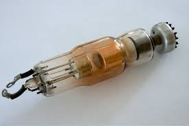
The key parameters of integrated X-ray sources include focal spot size, voltage, current, power, target material, cooling method, radiation angle, and window material/thickness. These parameters are interrelated and collectively determine the source’s performance and application range. Consider these factors based on your specific needs when selecting and using an integrated X-ray source.
1.Focal Spot Size
The focal spot size refers to the small area where the electron beam impacts the anode (target material) inside the X-ray tube. It directly affects the size of the X-ray beam spot and the spatial resolution of the image.
A smaller focal spot size produces a finer X-ray beam spot, which improves the spatial resolution of the image, making details clearer. Fine focal spots provide more precise detail representation, making them suitable for high-resolution applications such as precision industrial inspection or high-resolution medical imaging. As the power of the X-ray source increases (i.e., with higher acceleration current or voltage), the focal spot size typically enlarges. This is due to the concentration of heat on the target material at high power, causing thermal expansion and material characteristics to enlarge the focal area. Therefore, high-power applications may require a balance between focal spot size and thermal management.
2.Voltage
Voltage is the electric potential difference applied between the cathode and the anode (target material) in the X-ray source. It is a key parameter for generating X-rays. The tube voltage directly determines the energy level of the X-rays.
Higher voltage results in higher energy X-rays. High-energy X-rays have shorter wavelengths and can penetrate thicker materials, making them suitable for inspecting denser or thicker substances. This is particularly important in industrial inspection and medical imaging. However, excessive voltage can lead to image overexposure, causing loss of detail or reduced image contrast. High power at high voltage can increase the focal spot size, affecting spatial resolution. Excessive voltage may reduce image resolution, especially in applications requiring fine structural details. High voltage operation can also impact the stability of the X-ray source and potentially shorten the device’s lifespan, necessitating effective cooling and maintenance measures.
3.Current
Current is the flow intensity of the electron beam in the X-ray source, usually measured in milliamps (mA). It is a key parameter that determines the intensity of the X-rays produced.
The size of the current directly affects the intensity of the X-rays. Higher current can generate more X-ray particles, thereby increasing imaging sensitivity and efficiency. This is particularly important in applications requiring rapid imaging or high-intensity X-rays. A higher current can reduce imaging time and improve the efficiency of image acquisition. In situations requiring rapid scanning or large-area inspection, a higher current accelerates the image capture process. However, excessive current can cause overheating of the X-ray source, increasing the thermal load on the equipment. Prolonged operation at high current levels may accelerate device aging and damage. Overheating can affect the stability of the equipment, leading to unstable operation or failures. Therefore, an effective cooling system is necessary to maintain the equipment’s normal operating temperature. Instability in current can lead to fluctuations in X-ray intensity, affecting imaging stability and quality. In high-demand imaging applications, such fluctuations can impact the accuracy and consistency of the final image.
4.Power
Power is an important parameter of the X-ray source, reflecting its output capability. It is the product of tube voltage and tube current, given by the equation Power=Voltage×Current, and is typically measured in watts (W).The level of power directly determines the output strength and penetration capability of the X-ray source. Higher power enables the X-ray beam to penetrate thicker materials, providing higher imaging quality and detailed internal information in applications such as industrial inspection and medical imaging.
A high-power X-ray source can complete scanning tasks more quickly, reducing imaging time. This is particularly important in applications requiring rapid inspection or real-time monitoring. Higher power can improve image quality, making it clearer and more detailed. However, excessive power may increase the focal spot size, which can affect the spatial resolution of the image. Achieving maximum power at the smallest focal spot size may be impractical. As the power on the target increases, the focal spot size typically enlarges, which can lead to reduced image resolution. Therefore, it is necessary to find a balance between power and focal spot size to meet specific imaging requirements. High-power operation generates significant heat, requiring an effective cooling system to prevent equipment overheating, ensure stability, and extend the equipment’s lifespan.
5.Other Key Parameters
Target Material
The target material is the substance in the X-ray source where the electron beam strikes to produce X-rays. Common target materials include tungsten, chromium, and copper.
X-ray Production Efficiency: The material properties of the target determine the efficiency of X-ray production. Tungsten is commonly used in high-power X-ray sources due to its high atomic number and excellent thermal conductivity.
X-ray Performance: The physical properties of different target materials (such as melting point, thermal conductivity, and X-ray emission characteristics) affect the quality of X-rays and the stability of the equipment. For example, tungsten maintains stable performance at high power levels, and its high melting point makes it more reliable under high-temperature conditions.
Cooling Method
The cooling method refers to the system used in integrated X-ray sources to manage the heat generated by the equipment. Common Cooling Methods is as below:
Air Cooling: Uses fans or airflow to dissipate heat, suitable for small to medium-power X-ray sources.
Oil Cooling: Uses oil as the cooling medium, suitable for high-power X-ray sources due to oil’s high heat capacity and good thermal management performance.
An effective cooling system maintains the equipment within a safe operating temperature range, preventing overheating and enhancing the stability and longevity of the equipment. Choosing the appropriate cooling method ensures the equipment operates normally under high loads and reduces performance degradation or equipment failure due to overheating.
Radiation Angle
The radiation angle refers to the range of angles at which the X-ray beam is emitted from the source. The radiation angle determines the coverage area of the X-ray beam. A larger radiation angle can cover a broader area, suitable for large-scale inspection or scanning. Different radiation angles are suited for different applications, such as small angles for detailed local inspections and large angles for overall scanning and broad-range detection.
Window Material/Thickness
The window is the channel through which X-rays exit the source, and its material and thickness affect X-ray transmission and image quality.
Material Choice: The selection of window materials impacts X-ray transmission. For example, lead glass or other high-density materials are often used for shielding and protection.
Thickness: The thickness of the window needs to balance between X-ray transmission and structural strength. A window that is too thick may reduce X-ray intensity, while a window that is too thin may not effectively protect the equipment and operators. These parameters affect the performance, imaging quality, and stability of the integrated X-ray source.
In summary, the key parameters of an integrated X-ray source include focal spot size, voltage, current, power, target material, cooling method, radiation angle, and window material/thickness. These parameters are interrelated and collectively determine the performance and application range of the X-ray source. When selecting and using an integrated X-ray source, it is essential to consider these parameters based on the specific application and requirements.


