What are X-rays?
X-rays are a form of electromagnetic radiation with high energy, allowing them to penetrate solid materials and living tissues. This makes them essential for applications like medical diagnostics and industrial testing.
Discovery and Historical Background of X-rays.
X-rays were discovered in 1895 by German scientist Wilhelm Conrad Röntgen, who named the radiation “X” to denote its unknown nature. Sometimes, X-rays are also referred to as Röntgen rays.
How Are X-rays Generated?
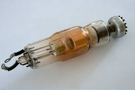
X-rays are generated in an X-ray tube, where a filament power supply heats the cathode, causing thermionic emission of electrons. A high voltage applied between the cathode and anode accelerates these electrons toward the anode. Upon collision, X-rays are generated. The X-ray tube, the heart of the x ray generating system, is maintained under vacuum to ensure efficient electron acceleration without interference from air molecules.
Properties of X-rays.
- X-rays have high energy, enabling them to penetrate the materials being examined.
- Their wavelength is shorter than visible light.
- X-rays produce ionizing radiation, which can be harmful to living tissues.
- X-rays are invisible to the human eye.
- X-rays are produced by applying high voltage to the X-ray tube, which accelerates electrons to generate radiation.
Types of X-rays.
X-rays are used in various medical applications:
- Plain X-ray: A 2D image created by passing a single X-ray beam through the body.
- Computed Tomography (CT): Combines multiple X-ray images taken from different angles to create cross-sectional views of the body.
- Mammography: Used specifically to examine breast tissue.
- Fluoroscopy: Provides real-time moving images of the inside of the body using X-rays.
- Angiography: Uses X-rays to visualize blood vessels and organs.
- Bone Density Scan (DEXA): Measures bone density, helping to detect fractures and prevent osteoporosis.
- Dental X-rays: Aid dentists in diagnosing and monitoring oral health.
In industry, X-rays are widely used for detecting internal defects, such as cracks or inconsistencies in materials, and for inspecting packaged foods and bulk food for foreign objects.
How X-rays Work?
X-rays are used in combination with detectors. As X-rays pass through objects, different materials absorb X-rays at varying levels depending on the material’s density and atomic number. The detector records the varying levels of attenuation to create an image. In medical imaging, bones absorb more X-rays than soft tissue, so bones appear white while soft tissues appear gray. In food inspection, X-rays are used to detect foreign objects inside food packages, as materials like metal, glass, or plastic absorb more X-rays than food, which helps identify contaminants.
Applications of X-rays
Medical Field
X-rays are widely used in medical diagnostics, such as detecting fractures, checking lung conditions, screening for cancer, and visualizing internal organs through technologies like CT scans. Dental X-rays are essential for diagnosing and monitoring oral health, especially before procedures like implants, periodontal surgery, or orthodontics.
Industrial Field
In the industrial sector, X-rays are commonly used for non-destructive testing (NDT) to detect internal defects, such as cracks or porosity in materials, without damaging the item being tested. They are also used to inspect semiconductor chips, circuit boards, and other components to ensure product quality.
Food Safety and Quality Control
X-rays are used in the food industry to detect foreign objects in food products without damaging the packaging or structure. This technology is vital for quality control and ensuring that products meet safety standards.
Security Inspection
X-rays are frequently used in security checks at airports, customs, and other high-security areas to scan luggage and cargo for prohibited or dangerous items.
Scientific Research
X-rays are used in various research applications, such as analyzing the chemical composition and elemental content of materials, examining ore, testing metal alloys, measuring coating thickness, and analyzing crystal structures.
X-ray Safety and Protection
X-ray radiation is ionizing, meaning it can be harmful to human tissue. To reduce risk, protective measures such as wearing lead aprons and thyroid collars, maintaining distance from the radiation source, and minimizing exposure time are essential. In industrial testing, shielding devices like lead curtains are commonly used to avoid direct exposure to the primary X-ray beam.
Trends in X-ray Technology
AI Integration:
Artificial intelligence is being used to enhance image analysis, identify anomalies, and assist radiologists in making quicker, more accurate diagnoses.
Miniaturization and Portability:
Advances in smaller, lighter, and more portable X-ray devices, improving space efficiency and mobility.
Radiation Dose Reduction:
Sophisticated software algorithms help minimize radiation exposure to patients and operators while maintaining high image quality.


